At the Hunterdon Breast Surgery Center we are your partner in making your breast health a priority. We provide many services to achieve that goal and we are committed to diagnosing, treating, and preventing problems that affect you and your breast health.
Microsite Admin Fields: Breast Surgery Center

Services
Services We Provide
Below are the biopsies that are performed at the Hunterdon Breast Surgery Center by our board certified breast surgeons:
Fine Needle Biopsy
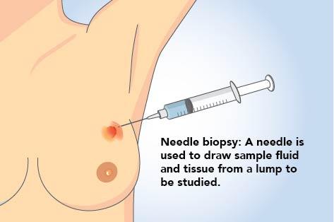
A fine needle biopsy of palpable lesions (lesions that can be felt) is the least invasive biopsy. It can be done in the doctor's office. Results are often available in 24 hours. During the procedure, a long, thin, hollow needle is placed in the palpable abnormality. The tissue is then sent off to pathology for analysis. This biopsy technique has the highest risk of a "false negative" — a biopsy result that says "normal," even though cancer is present. Therefore this is not as commonly performed.
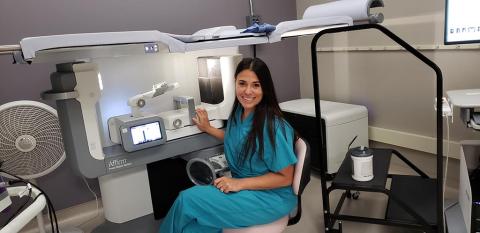
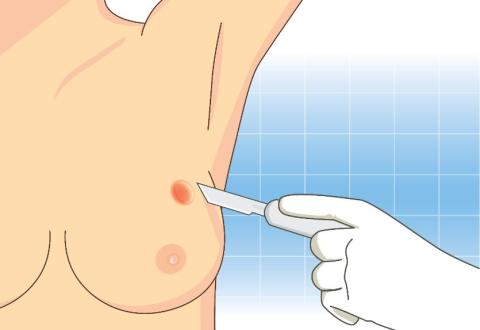
Stereostatic Biopsy
This type of biopsy uses mammograms to pinpoint the location of suspicious areas within the breast. For this procedure, you lie face down on a padded biopsy table with one of your breasts positioned in a hold in the table. You may need to remain in this position for 30 minutes to one hour. The table is raised several feet. The equipment used by your board certified breast surgeon is positioned beneath the table. Your breast is firmly compressed between two plates while mammograms are taken to show the radiologist the exact location of the area for biopsy. A small incision – about 4 millimeters – is made into your breast. The breast surgeon inserts either a needle or a vacuum-powered probe and removes several samples of tissue. The samples are sent to a laboratory for analysis. Results are usually reported within 5-7 days. At the Hunterdon Breast Surgery Center, the breast surgeons use a state-of-the-art stereotactic biopsy table that has 3D capability. This allows for the biopsy of abnormalities that might otherwise be missed with other technology. (The image on the left is our state-of-the-art stereotactic biopsy table where stereotactic biopsies are performed.)
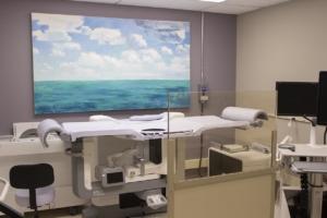
Ultrasound-Guided Needle Biopsy
This type of core needle biopsy involves ultrasound – an imaging method that uses high-frequency sound waves to produce precise images of structures within your body. During this procedure, you lie on your back on an ultrasound table. Using ultrasound, your breast surgeon locates the mass within your breast, makes a small incision to insert the needle and takes several core samples of tissue to be sent to a laboratory for analysis. (The image is our ultrasound table where ultrasound-guided biopsies are performed.)
Excisional Biopsy
Excisional biopsy is the most involved kind of biopsy. It attempts to remove the entire suspicious lump of tissue from the breast, in the operating room or surgery center. The purpose of this procedure is to make a diagnosis. Even if the lumpectomy takes out all of the cancer in the breast with clear margins, if breast cancer is diagnosed at that time, an additional surgery may be required.
MRI-Guided Core Needle Biopsy*
*The MRI-Guided Core Needle Biopsy is performed at Hunterdon Medical Center under the recommendation of your breast surgeon.
This type of core needle biopsy is done under the guidance of MRI – an imaging technique that captures multiple cross-sectional images of your breast and combines them using a computer, to generate detailed 3-D pictures. This biopsy is selected if the abnormality is only seen on MRI imaging. During this procedure, you lie face down on a padded scanning table. Your breasts fit into a hollow depression in the table. The MRI machine provides images that help determine the exact location for the biopsy. Several samples of tissue are taken and sent to a laboratory for analysis.

Surgical Procedures
Excisional Biopsy
Excisional biopsy is the most involved kind of biopsy. It attempts to remove the entire suspicious lump of tissue from the breast. This is the surest way to establish the diagnosis without missing the cancer tissue (winding up with a false negative). Both incisional and excisional biopsies can be done in an outpatient center or hospital, using anesthesia. The purpose of this procedure is to make a diagnosis. Even if the lumpectomy takes out all of the cancer in the breast with clear margins, if breast cancer is diagnosed, additional surgery may be required.
Lumpectomy/Partial Mastectomy
A Lumpectomy or Partial Mastectomy is a surgical procedure performed on a breast abnormality with a previous diagnosis by a needle biopsy. The suspicious lump and some surrounding tissue is excised and analyzed by pathology. Breast lump removal is generally completed as an outpatient surgery. You will be given a form of anesthesia. The procedure generally takes about 1 hour. The surgeon makes a small incision on your breast. The cancer and some of the normal breast tissue around it is removed. A pathologist examines a sample of the removed tissue to make sure all the cancer or abnormality has been taken out.
With a diagnosis of cancer, sometimes small metal clips will be placed inside the breast to mark the area of tissue removal. This makes the area easy to see on future mammograms. It also helps guide radiation therapy, when needed. The surgeon will close your skin with stitches, which are under the skin and dissolvable. Your doctor will send the “lump” to a laboratory for more testing.
Sentinel Lymph Node Biopsy
A Sentinel Lymph Node Biopsy is the removal and examination of the sentinel node(s) (the first lymph node(s) to which cancer cells are likely to spread from a breast tumor). To identify the sentinel lymph node(s), radiology will inject a radioactive tracer the day prior to surgery, that will travel to the first lymph node. Your breast surgeon will inject a blue dye in the breast, right before surgery, which will also travel to the first lymph node. The surgeon then uses a probe to find the sentinel lymph node(s) containing the radioactive substance and stained with dye. The surgeon then removes the sentinel node(s) to check for the presence of cancer cells, which will be sent to pathology for evaluation.
Axillary Dissection
An Axillary Dissection is a surgery to remove multiple lymph nodes found in the axilla (armpit) of the side of diagnosed breast cancer.
Mastectomy
A Mastectomy is a surgery to remove the entire breast. Most of the time, some of the skin and the nipple are also removed. The surgery is most often done to treat breast cancer. Before surgery begins, you will be given general anesthesia. This means you will be asleep and pain-free during surgery. There are different types of mastectomies. Which one your surgeon performs depends on the type of breast problem you have. Most of the time, mastectomy is done to treat cancer. However, it is sometimes done to prevent cancer (prophylactic mastectomy). The surgeon will decide which of these options are best for you, and will perform one of these operations:
- Nipple sparing mastectomy: The surgeon removes the entire breast but leaves the nipple and areola (the colored circle around the nipple) in place. If you have cancer, the surgeon may do a biopsy of lymph nodes in the underarm area to see if the cancer has spread. This is usually performed in conjunction with breast reconstruction, with a plastic surgeon (who specializes in breast reconstruction).
- Total or simple mastectomy: The surgeon removes the entire breast along with the nipple and areola. If you have cancer, the surgeon may do a biopsy of lymph nodes in the underarm area to see if the cancer has spread. This can be done with or without breast reconstruction.
- Modified radical mastectomy: The surgeon removes the entire breast with the nipple and areolar along with some of the lymph nodes underneath the arm. This can be done with or without breast reconstruction.
- Skin-sparing mastectomy: The surgeon removes the breast with the nipple and areola with minimal skin removal. If you have cancer, the surgeon may do a biopsy of lymph nodes in the underarm area to see if the cancer has spread. This can be done with or without breast reconstruction. One or two small plastic drains or tubes are very often left in your chest to remove extra fluid from where the breast tissue used to be.
A plastic surgeon may be able to begin reconstruction of the breast during the same operation. You may also choose to have breast reconstruction at a later time. If you have reconstruction, a skin or nipple sparing mastectomy may be an option.
Mastectomy generally takes about 2 to 4 hours, depending on the surgery selected.
Oncoplastic Surgery
Oncoplastic surgery is a surgical approach to treating breast cancer that can improve the cosmetic outcomes for many patients. The surgeons combine breast cancer tumor removal and plastic surgery techniques during the same surgical procedure. By doing this, patients can have their cancer removed while preserving the physical appearance of their natural breasts at the same time.

High Risk Screening
Here at the Hunterdon Breast Surgery Center, we offer appropriate surveillance and management for our patients that are at an elevated risk for developing breast cancer. If you wish to find out if you are at an elevated risk for developing breast cancer, please do not hesitate to schedule a consultation with one of our Board Certified Breast Surgeons.
If you are at high risk because of a strong family history of breast cancer or atypical cells found on previous biopsies, you should be under increased surveillance. After consultation with your breast surgeon, they will develop a screening plan tailored to your unique situation. Current recommended screening guidelines include:
- A monthly self-breast examination
- A biannual breast examination and evaluation by a breast surgeon (or another healthcare provider)
- A mammogram every year starting at 40 years old (or younger if family history of breast cancer at a young age)
- Breast MRI (magnetic resonance imaging) on an annual basis
- Ultrasound (usually in conjunction with an annual mammogram)
Genetic Testing
If you have a significant family history of cancer, evaluation from our genetic counselors and possibly genetic testing may be appropriate for you. After consultation with your breast surgeon, they may refer you for additional evaluation.
The Family Risk Assessment Program (FRAP) at Hunterdon Regional Cancer Center is designed to assist individuals with a personal or family history of cancer to understand how hereditary factors may contribute to cancer risk and how that risk can be reduced and/or managed. The main goals of a cancer genetic risk assessment are to collect and evaluate personal, medical, and family health information, to identify those high-risk individuals who may benefit from either genetic testing, cancer risk reduction interventions, or early detection strategies, and to improve the quantity and quality of life.
To find out more about your risk of developing breast cancer, depending on your family history and other associated risk factor, consultation with genetic counselors is recommended. For more information, please visit: Family Risk Assessment Program (FRAP) or call (908) 788-2535.

Here at the Hunterdon Breast Surgery Center, we offer partial breast radiation on patients diagnosed with breast cancer, when appropriate. Partial breast radiation is performed under the supervision of our radiation oncologists at the Hunterdon Regional Cancer Center. It is a type of breast radiation, which lasts 5 days instead of the usual 4-6 week course, and requires insertion of a temporary catheter, which is placed in the breast. The radiation catheter is inserted at the Hunterdon Breast Surgery Center by your breast surgeon. Appropriateness of partial breast radiation is decided between your breast surgeon and our radiation oncology team following surgery.
Additional Resources
The Hunterdon Breast Surgery Center works very closely with the Hunterdon Women’s Imaging Center. We conveniently share the same building on Route 31 in Flemington, New Jersey. In addition, Hunterdon Advanced Imaging Center, located in the Bridgewater Medical Office Building, and Hunterdon Advanced Imaging at Clinton, located in the Clinton Health Campus, both offer 3D mammography.
Mammogram Guidelines
Hunterdon Health recommends that women begin breast cancer screening at age 40. If there is a history of breast cancer in your immediate family (such as a mother, aunt, or sister), you may be considered high risk. Women at high risk – because of family history, a breast condition, or another reason – need to begin screening earlier and/or more often. Please discuss with your healthcare provider.
Please discuss with your breast surgeon or healthcare provider for further information and screening recommendations.
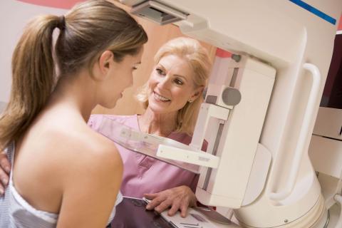
3-D Digital Mammography Services
A mammogram is a low-dose X-ray of the breast. It can detect a breast lump nearly two years before it can be felt. Digital mammography is the most advanced technology to date for detecting breast cancer. The digital mammography procedure is essentially the same as standard film mammography but uses a computer and digital image instead of film. Digital mammograms allow the image to be acquired and displayed immediately, reducing the time that the patient must remain still. This expedited process provides the patient with a more convenient and comfortable mammogram. In addition, a digital image can be enhanced and altered to be seen more clearly and to make a more accurate diagnosis. This image manipulation eliminates the need for a woman to repeat her mammogram if the first image is deemed unusable.
Screening mammograms evaluate breast health in women with no symptoms and are used for those who seek routine breast evaluation. Diagnostic mammograms are used to diagnose breast disease in women with symptoms of a breast problem: dimpling, or a change in the texture of the skin of the breast, a lump, or discharge from the nipple.
All of the diagnostic testing performed at our centers is done with the most advanced, state-of-the-art technology. Hunterdon Medical Center is accredited by the American College of Radiology as a nationally approved Mammography and Ultrasound Center. In addition, Hunterdon Medical Center is the only Breast Center in Hunterdon County to be accredited by the National Accreditation Program for Breast Centers.
Hunterdon Women's Imaging Center
Hunterdon Women’s Imaging Center features leading-edge technology in a tranquil environment. The comprehensive diagnostic services offered at this location include:
- 3D Mammography
- Breast Ultrasound
- Ultrasound
- Bone Densitometry (DEXA)
For more information, click here or call 908-237-4150 to schedule an appointment.
Advanced Imaging at Bridgewater
Advanced Imaging at Bridgewater is located at 1121 Route 22 West in Bridgewater, NJ. The comprehensive diagnostic services offered at this location include:
- 3D Mammography
- CT Scan
- MRI
- Bone Density Scan
- Ultrasound
- X-ray
For more information, click here or call 908-237-4150 to schedule an appointment.
Hunterdon Advanced Imaging at Clinton
Hunterdon Advanced Imaging at Clinton is located at the Clinton Health Campus, 1738 Route 31 North, Suite 108, in Clinton, NJ. The comprehensive diagnostic services offered at this location include:
- CT Scan
- Calcium Scoring Studies
- MRI (3T)
- X-ray (walk-in)
- 3D Mammography (walk-in)
- Ultrasound
- Bone Density Scan
For more information, click here or call 908-237-4150 to schedule an appointment.

At the Hunterdon Breast Surgery Center, we work closely with board-certified plastic surgeons to guarantee you the most natural outcome so that you can look and feel your best.
Breast Reconstruction
Breast Reconstruction is the re-creation of a woman’s breast through surgery, either with an implant or her body’s own tissue. This procedure can be done at the time of mastectomy (surgical removal of the breast) or delayed until you are medically and/or psychologically ready. The plastic surgeons we work with employ a variety of specialized techniques designed to rebuild the breast and restore its natural look. All procedures are preceded by an extensive consultation designed to answer all questions and discuss the most appropriate plan for you.
Implant-Based Breast Reconstruction
Implant-Based breast reconstruction is divided into two steps: tissue expansion and placement of a breast implant.
Tissue Expansion A tissue expander is a temporary implant placed under the muscle and tissue following a mastectomy. The temporary implant makes room for a permanent replacement implant of the same size.
Placement of a Breast Implant After the tissue has been expanded, the tissue expander is exchanged for a breast implant. In some patients, placement of the implant can be done at the time of mastectomy without the need for a tissue expander. There are two types of implants: saline-filled and silicone gel-filled.
Autologous Breast Reconstruction
Autologous breast reconstruction means reconstruction using tissue – skin, fat and sometimes muscle – from another place on the body. The plastic surgery term for a piece of tissue like this is called a flap.
PEDICLED FLAPS
If the blood supply to the flap remains attached, it is called a pedicled flap. There are two types of pedicled flaps:
- Latissimus Dorsi Flap
- Latissimus Dorsi is the name of a back muscle
- In this procedure, skin, fat, and muscle are moved from the back to re-create the breast
- This may be combined with a tissue expander and/or breast implant
- Pedicled TRAM Flap
- TRAM (transverse rectus abdominis muscle) is one of the muscles of the abdomen
- In this procedure, skin, fat, and muscle are moved from the abdomen to re-create the breast
- In some cases, a BARs (bony anchor reinforcement) TRAM Flap is performed to reduce the rate of post-operative hernia. This innovative technique uses reinforced mesh that is placed on the abdominal wall at the time of reconstruction
FREE FLAPS
If the blood supply to the flap is separated and reattached, it is called a free flap. To perform a free flap, your surgeon must have expertise in microsurgery, where the small blood vessels are reattached under a microscope. There are three types of free flaps:
- Free TRAM Flap
- TRAM (transverse rectus abdominis muscle) is one of the muscles of the abdomen
- In this procedure, skin, fat, and muscle are moved from the abdomen to re-create the breast
- DIEP Flap
- The DIEP (deep inferior epigastric artery) is the blood supply to the abdominal skin and fat
- In this procedure, skin and fat are moved from the abdomen to re-create the breast
- Non-abdominal Free Flap
- In certain patients, it may be possible or necessary to use tissue from another part of the body to re-create the breast
- These areas include the hips, buttocks and posterior thigh
Please discuss your plastic surgery options and the plastic surgeons affiliated with Hunterdon Medical Center with your breast surgeons. We will work together to provide you with the best plan to achieve the best outcome.
Make an appointment today!